Biomedical engineering is an interdisciplinary research area that utilizes engineering methods to solve medical problems. Approaches include conducting physical experiments, creating mathematical models, scouring existing literature, forming hypothesis, and making sense of research finding. While biomedical engineering can cover a wide range of topic areas, our work focuses on biomechanics of human movement, neuromuscular control, characterization of biological tissues, injury biomechanics, and the design of medical devices.
Correlating Brain Strain from Head Impacts to Cognitive Function in Contact Sports
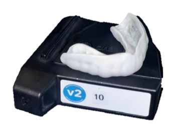
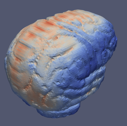
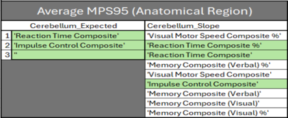
Traumatic Brain Injuries (TBIs) and mild TBIs (mTBIs) pose a significant risk in American football. Wearable sensors in instrumented mouthguards (iMGs) were used to collect skull movement data upon impact and finite element modeling was used to calculate the resulting brain strain. For impacts over 30Gs, which were considered severe in this study, cognitive function tests were conducted and compared to baseline test to determine if cognitive impairment had occurred. Data from the brain strain model indicated the regions of the brain that experienced strain and the cognitive test measured performance in a plethora of different functions (e.g., verbal memory, visual memory, reactions speed, etc.). This generated a multitude of data and correlations were made between the strain in brain areas and specific brain functions.
This research was performed in collaboration with Pennsylvania State University. Head impact data was collected at WCU by Clayton Bardall, MSET graduate student and WCU football player. Brain strain analysis for head impacts was performed at Penn State using their cloud-based FEA analysis software, the Brain Simulation Platform, developed by Dr. Reuben Kraft and Ritika Menghani. Preliminary result showed that 1) the maximum elemental strain was the best predictor of brain strain, 2) 30Gs of linear acceleration was sufficient to induce cognitive impairment which may be associated with mTBIs (90Gs is often associated with concussions), and 3) experimentally we found that brain strain in the ventricles and cerebellum were the best predictors of cognitive dysfunction (we do not know why).
By fitting collegiate football players with iMGs, we seek to enhance our understanding of TBIs and improve player safety for future generations. Reuben and I hope to continue this research in the future. Clayton and Ritika have both graduated!
We have received a lot of media attention for our research. See some of the articles and videos on our research group’s news page.


Development of the Neural Prostheses
Many Americans have paralysis caused by strokes, injuries, or degenerative diseases. While some people have complete paralysis, many have partial paralysis leaving them with limited function. The case that we are interested in studying with this device is people with muscle weakness that have insufficient neural stimulation to properly activate the leg muscles when walking.
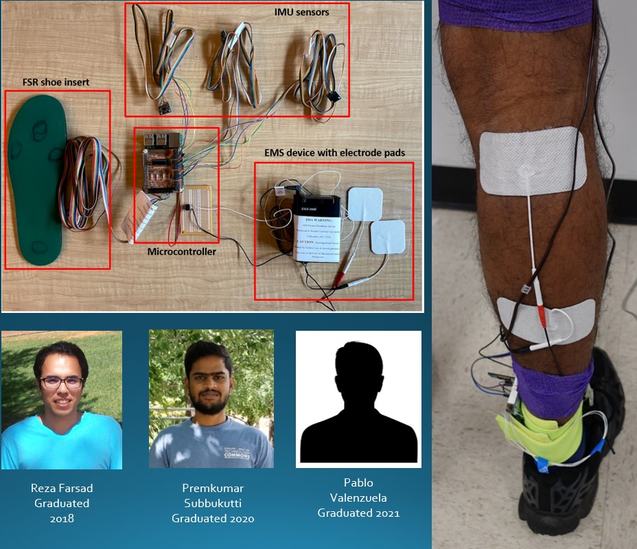
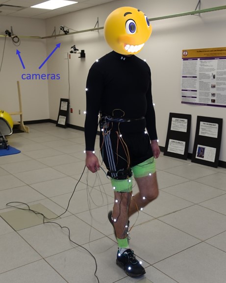
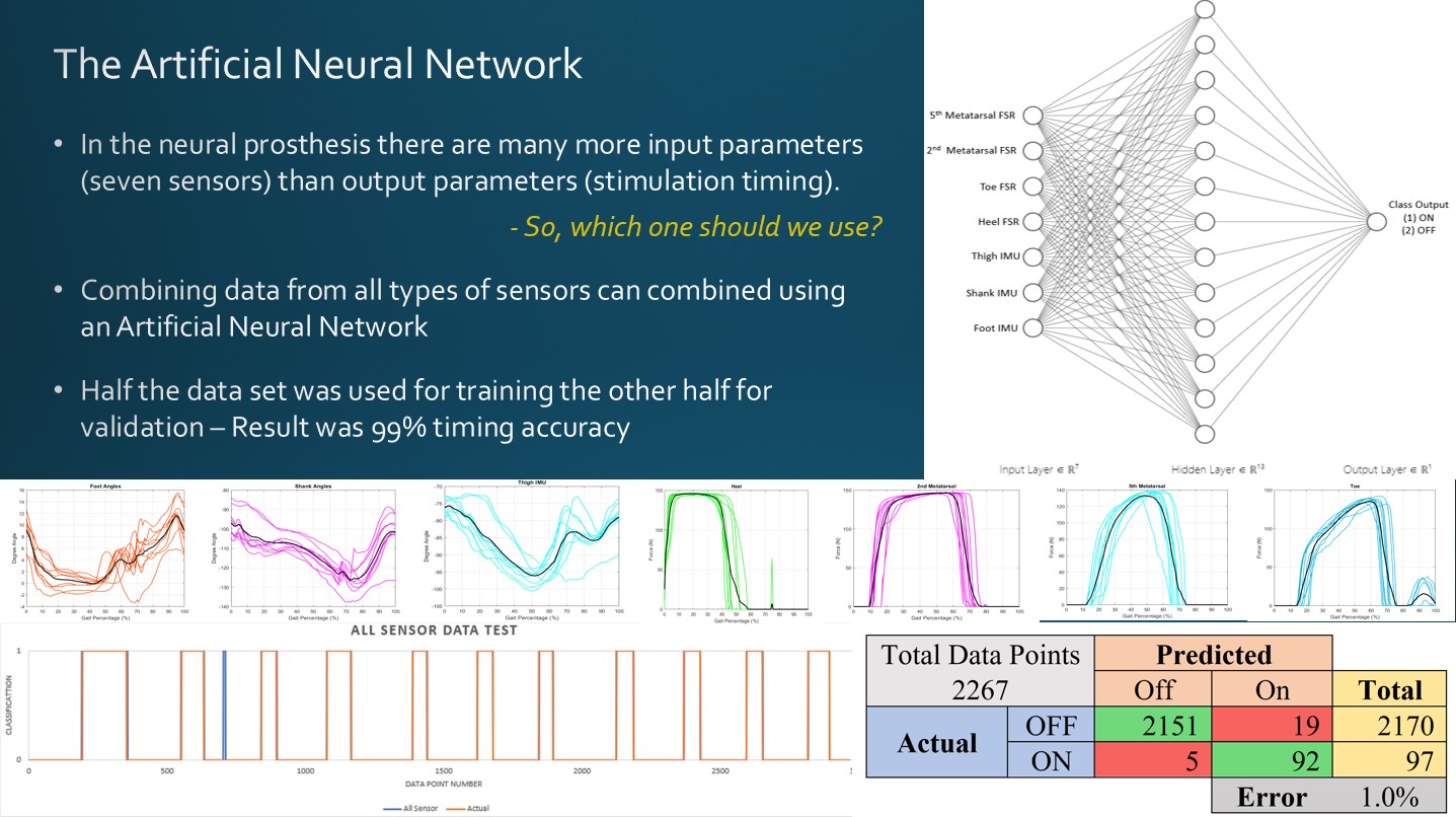
A prosthesis is “an artificial device to replace or augment a missing or impaired part of the body”. The neural prosthesis that we are developing is a limited risk investigational medical device approved for evaluation by the IRB at WCU. It utilizes a commercially manufactured functional electrical stimulator, IMUs with triaxial gyroscopes and accelerometers, foot pressure sensors, and a microcontroller. The device detects gait utilizing sensors on the leg and foot and uses a neural network to determine when to stimulate the muscles. Gait data was collected and half of it was used to train the neural network and the other half was used to test it. The test revealed that muscle stimulation during consistent normal walking had a 99% timing accuracy.
While these results are promising, additional work is needed to improve the robustness of the design and train the neural network for movement patterns different than normal walking.
Functional Electrical Stimulation Bicycle
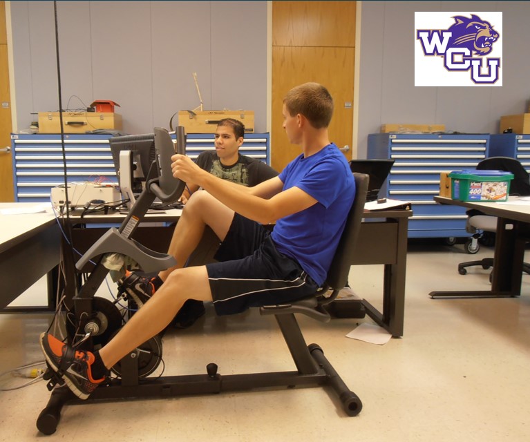
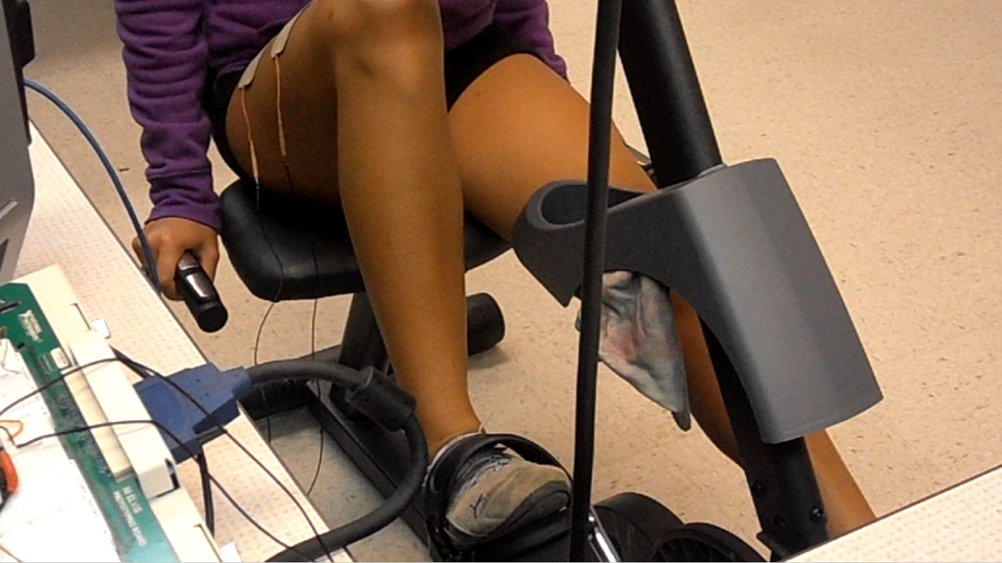
Our original electrical stimulation project was the FES bicycle. It used a recumbent bike that enabled a participant to cycle without worrying about falling. It also had the advantage that stimulation timing was controlled by the rotation of the foot pedals, so it was displacement controlled rather than timing controlled.
When the foot pedal was at a certain location in the pedal cycle, the stimulator would activate sending electrical impulses to artificially stimulate the muscle inducing a contraction.
The FES bicycle was designed to replace or augment the neural connection for spinal cord injury patients, to help rehabilitate stroke victims, or assist people with neurologically induced muscle weakness. In the case of a stroke, the FES bicycle has the potential to keep a patients muscles activated during the time that the brain is undergoing functional remodeling.
Like an astronaut exercising in space, the device reduces atrophy in the muscles and nerves during times when they may not normally be activated.
